Electron microscopy and calorimetry of proteins in supercooled
Por um escritor misterioso
Last updated 10 novembro 2024
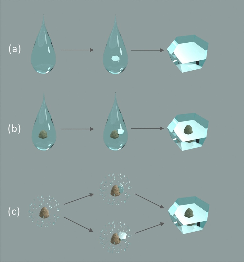

Structure of spruce budworm antifreeze protein.a, Stereoview of C

Types of differential scanning calorimeters: (a) heat-flux
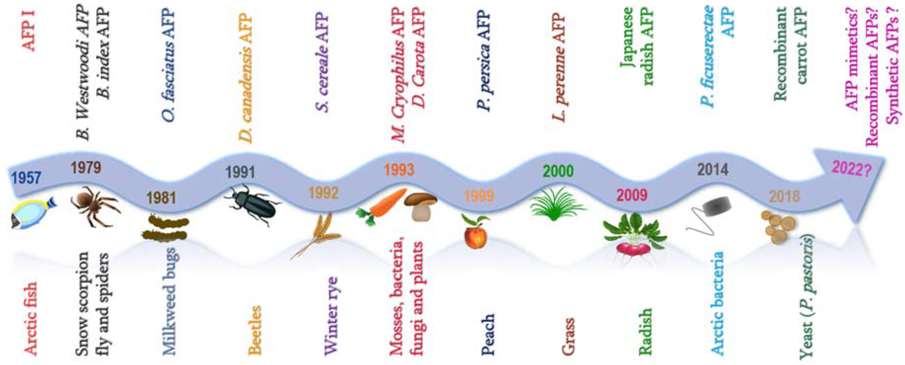
IJMS, Free Full-Text
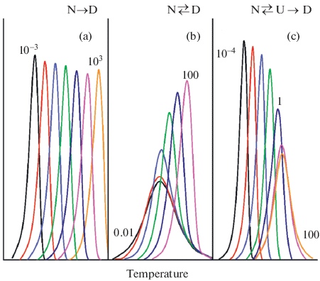
Fast Scanning Calorimetry of Organic Materials from Low Molecular
Structures of the investigated proteins and the tobacco mosaic
Stress treatments performed with apoferritin batch 1 (0.34 mg mL
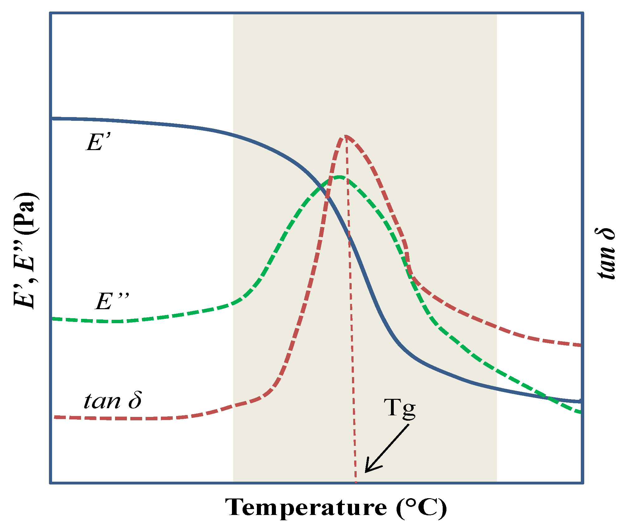
Polymers, Free Full-Text

PDF) Electron microscopy and calorimetry of proteins in
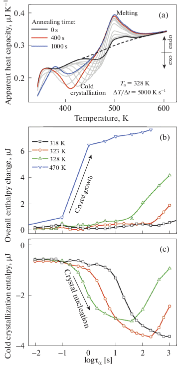
Fast Scanning Calorimetry of Organic Materials from Low Molecular

Supercooled Liquids and Glasses The Journal of Physical Chemistry

Vacuum-Induced Surface Freezing for the Freeze-Drying of the Human

a) Comparison of dielectric data (lines) and DDLS data (symbols
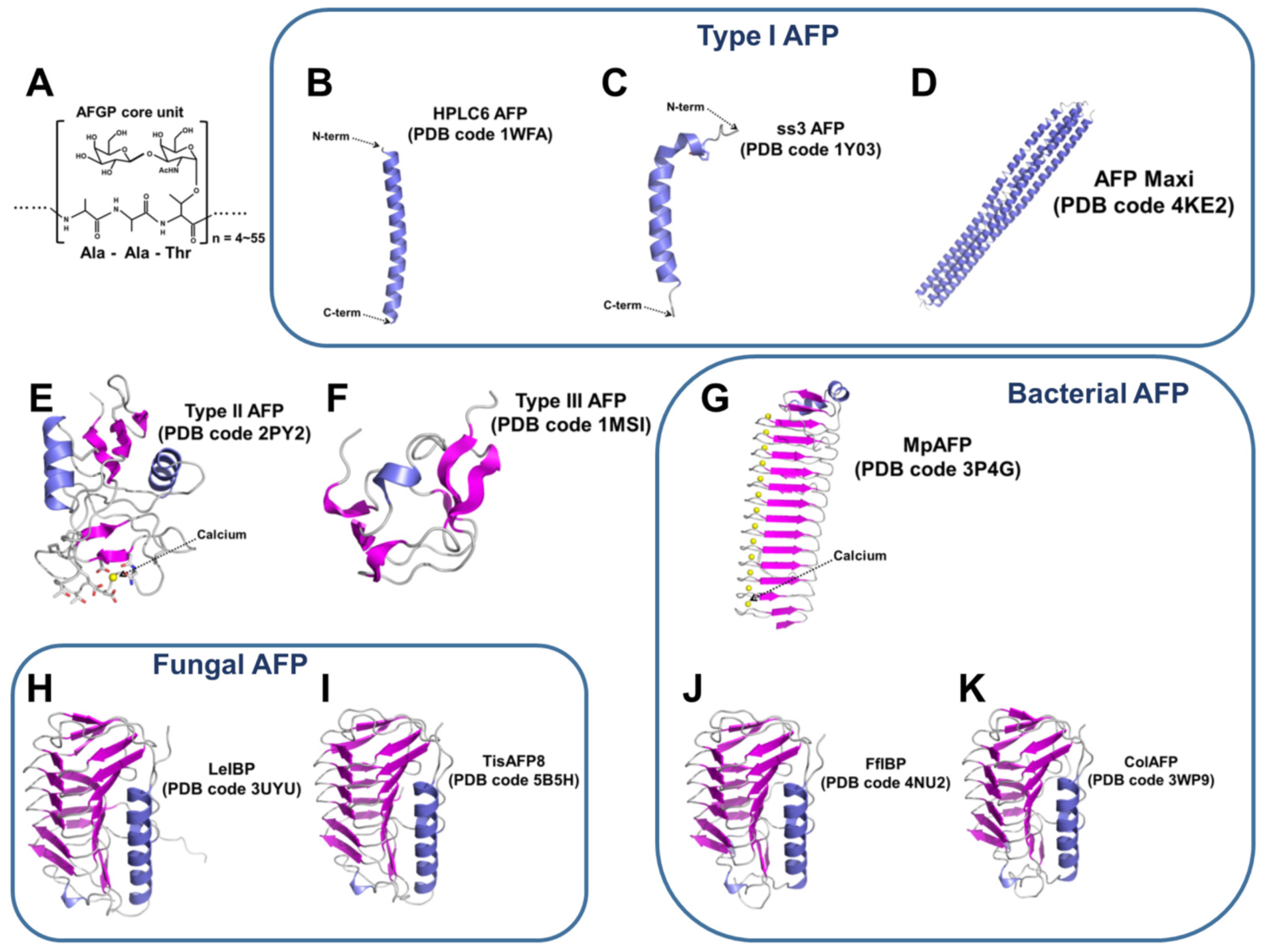
Marine Drugs, Free Full-Text

Role of Poloxamer 188 in Preventing Ice-Surface-Induced Protein

Temperature dependence of the relaxation times for e-PLL-water
Recomendado para você
-
 Shan♥️ on X: Prime bot is works like forex platform but in this10 novembro 2024
 -
 Join the CPR Training World Record & make history at WCA 2021 - WFSA10 novembro 2024
 -
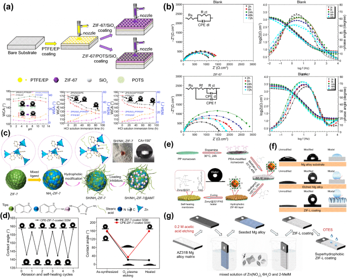 Recent progress of zeolitic imidazolate frameworks (ZIFs) in10 novembro 2024
 -
Guacathon 2021 LIVE: The 21st Birthday Celebration for Joaquin10 novembro 2024
-
 Local Business Sells Out of Wood Using Messenger Bots Case Study10 novembro 2024
 -
Video: GeT_RiGhT dies to a bot and f0rest can't stop laughing10 novembro 2024
-
 how to play with bots in call of duty 2|TikTok Search10 novembro 2024
 -
Discovery One - Truly exceptional! Meet the top 16 of the MLBB teams and join them on their exciting journey! Watch the live streams exclusively on Discovery One Facebook page, channel10 novembro 2024
-
 Water soluble calcium made from egg shells〡Third Insight Design and Nursery & nursery10 novembro 2024
 -
 News Briefs Hospitality Technology10 novembro 2024
você pode gostar
-
 Asterisk Light Novel Volume 12, Gakusen Toshi Asterisk Wiki10 novembro 2024
 -
 MOD MENU ROBLOX!! O MELHOR MOD ATUALIZADO COM ROBUX INFINITO E VÁRIAS FUNÇÕES!!10 novembro 2024
 -
 blaze and the monster machines lunch box10 novembro 2024
 -
kaguya sama temporada 3 capitulo 5|Búsqueda de TikTok10 novembro 2024
-
 Comentando: Hunter x Hunter – Episódio 148 – Final10 novembro 2024
 -
 Blountstown FL Basketball - FCA Sports > Home10 novembro 2024
 -
 Tensai Ouji no Akaji Kokka Saisei Jutsu Dublado - Episódio 2 - Animes Online10 novembro 2024
 -
 Akatsuki RAP Anime RAP AniBeat #SPECIALNARUTO10 novembro 2024
 -
 Aliceliese Lou Nebulis IX, KimiSen Wiki10 novembro 2024
 -
 Miquella and Malenia: The Full Story10 novembro 2024


